
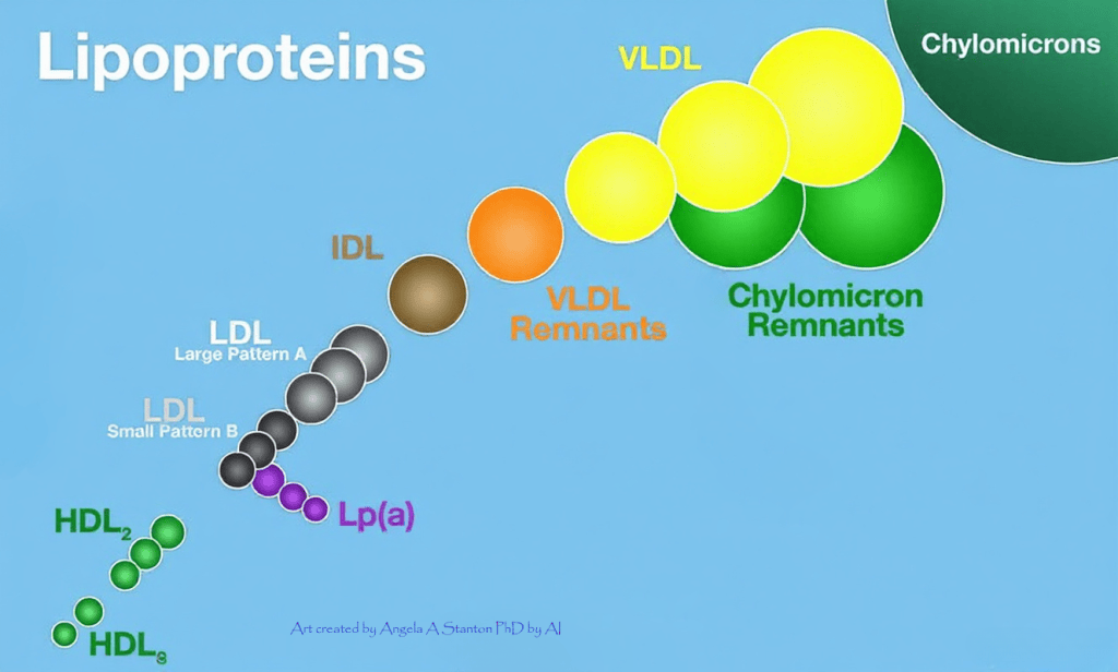
Lowering LP(a) with Medication (Pelacarsen) Failed in Clinical Trial!
Pelacarsen lowered Lp(a) but did not reduce the trial’s primary cardiovascular endpoint in the study population. While details are still hidden, to me, this means that Pelacarsen failed to protect against cardiovascular events despite lowering Lp(a).
Novartis today announced that the pelacarsen phase III Lp(a) HORIZON trial, a pioneering cardiovascular outcomes study, did not meet its primary endpoint of reducing the risk of cardiovascular events, a composite of cardiovascular death, non-fatal myocardial infarction, non-fatal stroke, and urgent coronary revascularization requiring hospitalization, compared to placebo in the overall study population. Read the original announcement by Novartis here.
The drug Pelacarsen, designed to sharply lower levels of a lipoprotein particle — known as lipoprotein(a), or “Lp(a)” — that scientific research has linked to heart disease. But in a multiyear Phase 3 study of more than 8,000 participants, a combination of Pelacarsen and standard medications didn’t lower the risk of major cardiovascular events when compared to those typical therapies and a placebo.
What is Lp(a)?
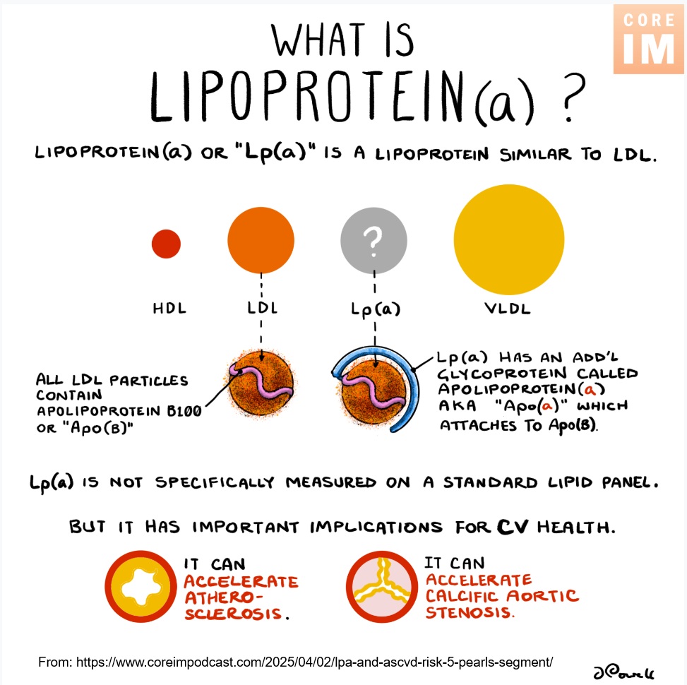
Lipoprotein(a) abbreviated as Lp(a), is a specific type of particle made of fat and protein that carries cholesterol through your bloodstream. It is similar to LDL, the so called “bad” cholesterol, but it contains an extra, sticky protein that makes it more likely to cause inflammation, build up plaque in your arteries, and form blood clots—or, at least, this is the current understanding.
Unfortunately, everywhere you look, the typical answer you get about cholesterol, including Lp(a) is that if you have high numbers of it (measured by blood test), you are on your way to or already have cardiovascular disease, atherosclerosis, and as soon as tomorrow you may be up for a heart attack or stroke. Of course, none of this is true. High cholesterol has long been linked to (note: *not causing*) a higher risk of heart and cardiovascular disease, making treatments that can reduce (or prevent) them mainstays of today’s cardiovascular care.
Genetic and epidemiological studies have shown a tie between Lp(a) and heart disease, basically based on the same principles. According to the American Heart Association, about 20% of people have high levels of Lp(a) and, as a result, are more likely to have harmful plaques accumulate in their arteries. But they can’t lean on diet or exercise to alter their Lp(a) counts since it is set by genetics.
Several large pharmaceutical companies have been racing to address the LP(a) problem by bringing forth a new crop of therapies able to plunge Lp(a) levels in one way or another. Pelacarsen was the first among these, making Novartis and Ionis’ trial one of the most eagerly anticipated in cardiovascular medicine.
There is an estimated $6 billion in U.S. sales per annum alone if a drug were to succeed in clinical testing.
High Lp(a) = You’re On Your Way to CVD?
The negative results revealed Friday raise questions not only about this approach on cardiovascular health, but also whether Lp(a) is truly an important genetic marker of a higher risk of heart and cardiovascular disease, or is it something that is “just there”.
The interesting thing is how companies tend to dig deeper and deeper into potential territories that make no sense at all and will likely end up changing the outcome expectations, lowering them, just so they can sell the drugs they created so they don’t go bankrupt. In my keyword search of PubMed Central, 836 academic articles popped up for the search “”Lipoprotein(a)” [Title] AND “causal” [All Fields]”.
Facts About Cholesterol
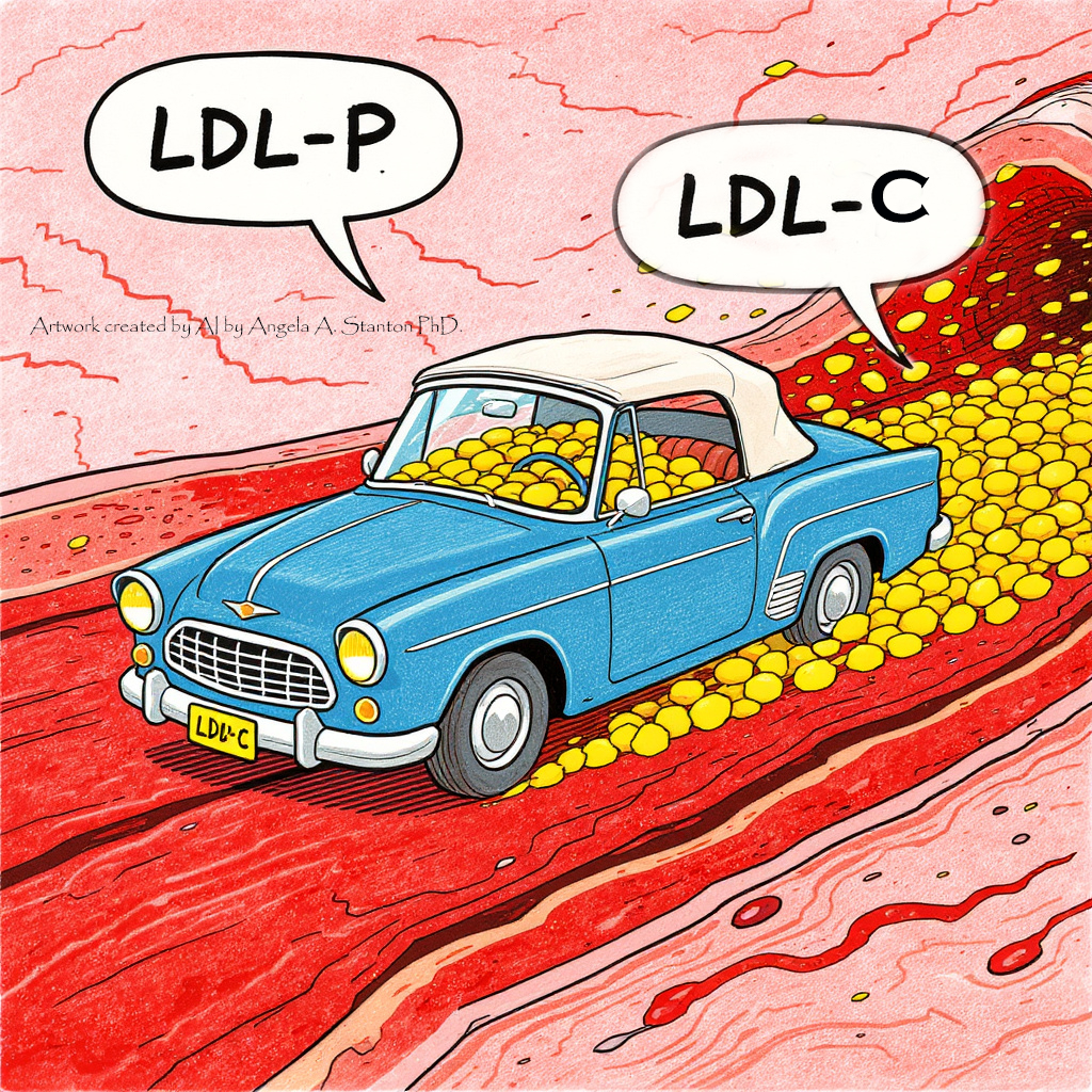
You often hear LDL, LDL-P, and LDL-C—all supposed to represent the so called “bad cholesterol”. So what exactly is it?
I always thought of LDL-P (P for Particle) as a badly generated term. A “particle” (to me) refers to a “single solitary unit”. The Cambridge Dictionary defines “particle” as “A tiny fragment or speck of matter, such as a dust or sand.” So it is naturally confusing when I discovered that the LDL “particle” has stuff in it, such as cholesterol and other things. 😉 This was one of the most confusing things in my understanding of cholesterol because as a mathematician, a particle means “one thing that has nothing in it”.
Here is a table to understand the rest of the terms in a simple blood cholesterol test:
| Measurement | What it tells you |
| LDL-P | The number of LDL particles circulating in the blood. |
| LDL-C | The amount (mass) of cholesterol carried by all those LDL particles combined. |
| ApoB | The number of ApoB molecules in the blood. Because each LDL, VLDL, IDL and Lp(a) particle carries one ApoB molecule, ApoB approximates the total number of these ApoB-containing particles, not just LDL particles. |
| Triglycerides (TG) | The amount of triglyceride carried inside circulating lipoprotein particles. In fasting blood, much of it is carried in VLDL; after eating, chylomicrons carry much of the dietary TG. |
| Fat-soluble vitamins A, D, E, K | These travel through the blood associated with lipoproteins and/or specific carrier proteins, depending on the vitamin and its chemical form. A routine vitamin blood test measures the vitamin concentration in serum/plasma, not the amount specifically inside LDL. |
In short: LDL-P counts the LDL particles. LDL-C measures how much cholesterol those LDL particles collectively carry. ApoB approximately counts all ApoB-containing particles, including LDL, VLDL, IDL and Lp(a). Triglycerides measure the amount of triglyceride being transported within circulating lipoproteins, rather than “free-floating fat” in the blood. Fat-soluble vitamins are also transported through blood using lipoproteins and/or specialized carrier proteins, depending on the vitamin.
Facts About Plaques
Let’s understand the anatomy of a plaque and go from there. Everywhere you check, you read that the plaque is in the arteries, it blocks the blood flow in the arteries, it is made of fat and cholesterol, eating saturated fat causes plaques, high cholesterol causes plaques, etc. Let’s learn what a plaque really is.
Where is the plaque?
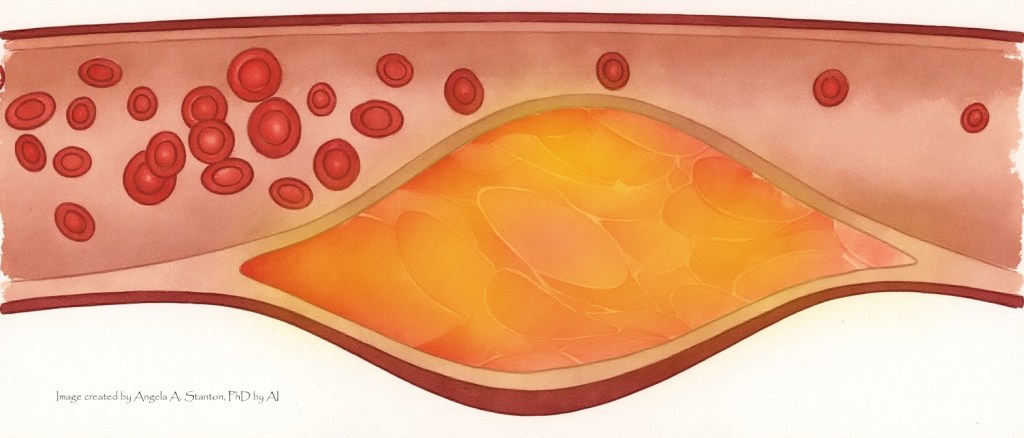
The plaque is not inside your arteries where the blood flows. It is in the wall of your arteries. Your arteries are made of a triple-wall muscular tissue plus the endothelium.
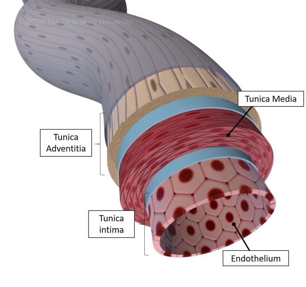
The location of the plaque is often misidentified in order to create a bigger drama. You usually see the plaque drawn in the inside of the artery—as in this picture taken from WebMD:
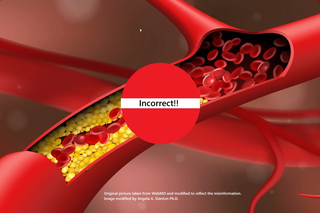
But this is incorrect. There are no plaques inside your arteries where the blood actually flows. The correct location of the plaque is actually inside the walls of your artery:
The very deep inside your blood vessels and arteries is the endothelium, a single-cell layer that is coated with glycocalyx. As you can see in the picture below, this glycocalyx looks somewhat like a bad hair day.
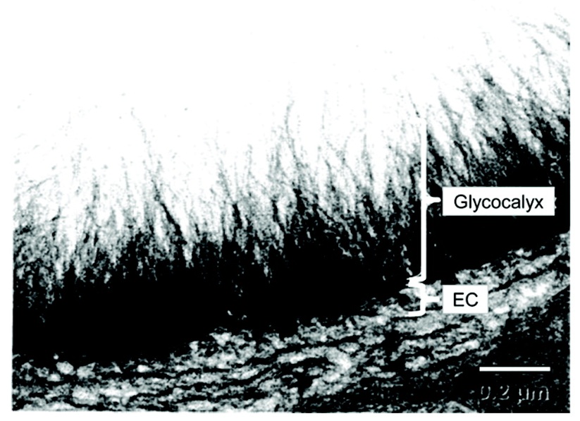
The image above is a transmission electron micrograph of a goat coronary capillary taken from here. It is a slippery layer, similar to how it also coats many fish, making them too slippery to hold. The glycocalyx forms a remarkably important interface between flowing blood and the endothelial cells beneath it. It regulates vascular permeability, mechanosensing, coagulation, inflammation, and interactions between circulating molecules and the endothelium. With your glycocalyx healthy and intact, only “controlled” elements can cross the glycocalyx and enter the endothelium in specific amount and with specific speed.
The glycocalyx is an important part of the vascular barrier specifically regulating interactions between the circulating lipoproteins and the endothelium. LDL-P can cross an intact endothelial layer through regulated transcytosis, so LDL-P entry does not explain plaque formation, since it happens naturally without forming plaques. The problem is the endothelial/glycocalyx dysfunction, which can be caused or aggravated by hyperglycemia, and which alters the permeability, transport speed and amount, inflammatory signaling, and the vascular homeostasis.
The pathological issue is when the ApoB-containing lipoproteins become retained at a larger rate and accumulate within the arterial intima, provoking a chronic inflammatory/remodeling response. And so the damage to the glycocalyx can cause a chain-reaction of events leading to the inflammatory response and the formation of the plaque. There are several models by which the crossing is facilitated more readily as a result of inflammation and damage.
Studies show that hyperglycemia and the enzymatic glycocalyx degradation that follows it independently increase LDL-P transport across the endothelial barrier and its retention in the arterial wall in various ways. This study shows that glucose, and specifically type 2 diabetes causes increased transport; this study looks at AGES, a downstream product of hyperglycemia, showing the process how LDL transport is increased in hyperglycemia, and this study shows that the uptake and transcytosis rates of the glycated (damaged by glucose) low-density lipoprotein (G-LDL) had a much higher rate of transcytosis than the healthy non-glycated LDL (N-LDL).
There are several other papers, each explaining different ways the transport is increased when hyperglycemia is present. A 42% glycocalyx degradation by hyaluronidase increased water flux velocity, LDL coverage, and mean maximum infiltration distance in isolated rat aortas (see here). Another study shows that a thinner glycocalyx at atheroprone carotid sinus regions (2.2 µm vs. 4.3 µm) produced a 2–3-fold increase in intimal LDL accumulation versus protected regions. The “last nail” in the coffin of the importance of hyperglycemia in plaque formation in several studies show that in healthy volunteers, even acute hyperglycemia (short time increase of glucose) reduced glycocalyx volume by ~50% within 6 hours, with shedding of hyaluronan and heparan sulfate into plasma (see these papers here, here, and here).
What is the plaque made of?

Plaque contains lipids and cholesterol, macrophages and other immune cells, smooth-muscle cells, connective tissue, calcium, cellular debris and, particularly in complicated lesions, thrombotic material. Once ApoB-containing lipoproteins become retained and modified within the intima, immune cells enter the area and macrophages take up lipid, producing foam cells. Smooth-muscle cells and extracellular matrix contribute to the fibrous cap, which can stabilize the lesion. Calcification and thrombosis are more complicated still.
In other words, much of what we call “plaque” isn’t simply a pile of cholesterol—it is the product of an ongoing inflammatory, repair and remodeling response to something happening inside the arterial wall.
So here is the question I think deserves considerably more attention: if hyperglycemia can damage the glycocalyx, increase endothelial LDL transport and promote intimal retention, how much of what we attribute to circulating LDL or Lp(a) is actually dependent upon the condition of the vascular barrier through which those particles must travel?
Back to Lp(a), Heart & CVD
I hope you can see having read the previous section about the inflammation that high blood glucose causes to the glycocalyx, allowing cholesterol to cross the endothelium in an unregulated dysfunctional inflammation superhighway, way easier and in larger numbers than normal, and then generate plaques.
My hypothesis is considerably more radical than the conventional reader may think: perhaps the concentration of cholesterol or Lp(a) is much less important than we have been led to believe. I think that what matters far more is whether ApoB-containing particles are moving through a healthy, regulated endothelial barrier or through a metabolically damaged, inflammatory one.
The failure of Pelacarsen doesn’t prove my hypothesis, but if dramatically lowering the presumed culprit fails to significantly reduce cardiovascular events, we should at least be willing to ask whether we have assigned the culprit too much importance.
In my opinion, the problem is the dysregulated transport, which is caused by chronic hyperglycemia. I see cardiovascular disease, heart attack, and stroke as cardiometabolic in origin. Cardiometabolic syndrome refers to the close connection between the heart and blood vessels, and the chemical processes that turn food into energy, the metabolic processes.
Might this be the reason why this drug failed?
In my opinion: yes. If the cause of heart attacks and cardiovascular disease is not Lp(a) or LDL-P, then reducing these won’t solve the problem.
Concluding Thoughts
In this article I do not recommend that you don’t test your cholesterol and don’t follow the recommendations of your doctors.
What I recommend is that you change your diet to reduce the glucose load because your cardiovascular system is ill-equipped to handling hyperglycemia.
Comments are welcomed, as always, and are moderated for appropriateness.
Angela


The glycocalyx is largely missing in the cholesterol conversation. Thank you Angela for highlighting it in your blog.
Without cholesterol or fat we die.
LikeLiked by 1 person
Indeed! We do die not only without cholesterol but just with low cholesterol. All of our immune system supporting fat-soluble vitamins travel in LDL. Reducing LDL is one of the biggest mistakes humans have ever dreamt up.
LikeLiked by 1 person
Gaslighting cholesterol for what exogenous sugar dysregulated is what clueless doctors do. Perpetual grazing on farinaceous kibble, “snacking”, keeps the body in a near-continuous post-prandial state voiding restorative mechanisms. Additionally, a lack of sunlight, or absent that D3/K2, is a complicating factor.
LikeLiked by 1 person
Dear Mani,
Thanks for your note. I hope you read my article and know that it is not gaslighting cholesterol for what hyperglycemia causes. Big pharma does.
And while the lack of sunlight is a complicating factor, most people I know supplement D3/K2–in fact, too many take too much and end up with toxic levels.
Toxic levels of D3 move calcium out of the bone instead of into it, and can participate in creating cardiovascular inflammation by flooding the blood with calcium.
I can’t tell you how often I see high serum calcium in a blood test! And that’s a recipe for atherosclerosis as well.
So general advice to take D3 and K2 can easily cause the very thing we are trying to prevent.
Angela
LikeLike
Pingback: The End of the Mediterranean Diet | Clueless Doctors & Scientists
Confirming your hypothesis:
https://www.uclahealth.org/news/article/healthy-blood-glucose-level-heart-disease-risk
LikeLiked by 1 person
Thanks Leonid. There are some issues with what they suggest. They write: “They highlighted a 2012 study by researchers in Israel that showed double the cardiovascular risk in individuals with fasting plasma glucose readings of 95-99 mg/dL, compared with values below 80 mg/dL. A study in 2019 by researchers in Denmark and Michigan revealed that the risk was also higher with increased variability within “normal” levels of HbA1c (a three-month average of blood sugar).”
I completely agree with this sentence: “Research has shown that blood sugar readings that fall just below prediabetes – on the higher side of “normal” – are associated with a notable increased risk of cardiovascular disease.”
They highlight “a 2012 study by researchers in Israel that showed double the cardiovascular risk in individuals with fasting plasma glucose readings of 95-99 mg/dL, compared with values below 80 mg/dL.”
But this is facing the same error I explained in my earlier response to your other question. As long as they only measure blood glucose, they will be greatly misinformed, becuase insulin compensates for a failing metabolic system. So having too low blood glucose is also a sign of problem, unless they are on the ketogenic diet. On the ketogenic diet the level of glucose in the blood can drop to very low since most of the body uses ketones. But in any other way of eating, where insulin is active, a very low blood glucose is an eye opener for possible sugar crash.
They are also considering the A1c a useful marker, which I don’t. The A1c assumes that, on average, a person’s red blood cell live for 3 months.
However, on the carnivore and the ketogenic diets the red blood cells tend to live to 4 months or even beyond. A1c looks at the “glycosylation damage” of the hemoglobin in the red blood cells–that is damage caused by glucose in the blood. But a red blood cell that lives for 4 months instead of 3, lives over 25% longer life than the red blood cell that lives for 3 months! A significantly more glycosylation damage will accrue to the red blood cells that live for 4 months instead of 3, and so the A1c will label people of keto and carnivore prediabetic, when, in fact, they are not.
So there are many variables here and most doctors and researchers do not follow all of them at once.
Angela
LikeLike
What is a healthy blood glucose level? Heart risk persists even
LikeLiked by 1 person
Dear Leonid,
A healthy blood glucose level is mostly between 80-100 mg/dL (4.45 – 5.55 mmol/L), with maximum increase from any meal being 20 mg/dL (1.1 mmol/L). I wear a CGM and when I sleep, my blood glucose is flat-lines between 80 and 90 mg/dL and my post meal blood glucose is usually between 100 – 115 mg/dL.
In terms of health risk persisting even then: yes, because how high your blood glucose goes is a factor of your metabolic health but also your insulin. Let me give you an example.
If you eat a lot of carbs in your day–normal SAD way of eating–your blood glucose most likely won’t go very high because your insulin is always very actively present in your blood.
So if you eat a lot of carbs, eating a corn on the cob will not spike your glucose much–at least for the first 20-30 years of your life–because your pancreas can release massive amounts of insulin on a dime. So your blood glucose will return to normal very fast and may even go slightly below what it was before eating the corn. This is referred to as reactive hypoglycemia. Reactive hypoglycemia is a marker for metabolic disease.
Another person may eat carnivore or ketogenic–I eat carnivorish–and so my insulin is not activated by carbs because I don’t eat any or very minimal. My blood glucose and insulin will behave very differently from yours, but it is also very healthy. In my case, eating a corn on the cob will allow my blood glucose to spike much higher and much faster than yours, because my insulin is “sleeping” so to speak. It is not used to being spiked. When the insulin finally releases, it will within minutes, the blood glucose will drop back to normal very fast, but never below what it was before eating, so there will be no reactive hypoglycemia.
Most doctors just give a blood glucose test, and if that comes our normal, they assume you are healthy. But had they also checked your fasting blood insulin, they may find you are not healthy at all.
In terms of the heart risk:
As long as you are metabolically healthy by eating right–no sugar, no ultraprocessed foods, no seed oils, no tropical fruits, fruit juices, smoothies and shakes with fruits, eating not much fruits at all, no starches or grains, you should be fine as long as you eat plenty of protein and fat. Only protein and fat are essential to eat. Carbs are not.
Good luck!
Angela
LikeLike